If You Viewed One Single Protein Using a Microscope
Using atomic force microscope it is possible to measure viscoelasticity of a single protein wherein its dissipative and elastic nature is directly and independently measured. Despite this limitation researchers have used combined single-molecule AFM and fluorescence-based approaches to track force-induced protein unfolding Sarkar Robertson.

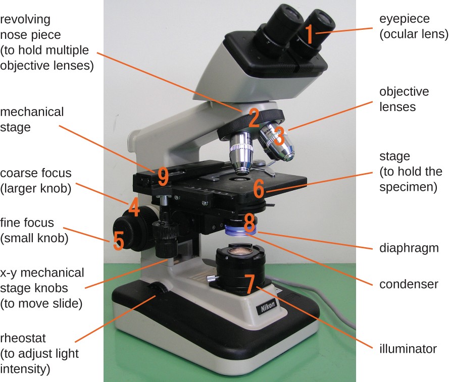
Instruments Of Microscopy Microbiology
It is now possible to create in a thin inorganic membrane a single sub-nanometer-diameter pore ie a sub-nanopore about the size of an amino acid residue.

. During these DNA interactions the search dimensionality is lowered from a three-dimensional diffusion process to a one. One side of the protein is anchored to a gold surface that acts as an electrode and. Typical field of view showing single.
Two antiparallel strands of DNA combine to form a double helix. If you viewed one single protein using a microscope you would observe multiple _____ structures. A Fluorescence photon burst event of single β-Galactosidase in 110.
He has reported protein images as small as 70 kDa. The aim of the measurement is to obtain a saturating concentration of MG-ester. Super-Resolution Microscopy and Single-Protein Tracking in Live Bacteria Using a Genetically Encoded Photostable Fluoromodule.
The specific interactions that permits this phenomenon occur by way of _____ bonds between _____. It is now possible to create in a thin inorganic membrane a single sub-nanometer-diameter pore ie a sub-nanopore about the size of an amino acid residue. New Light Microscope Can View Protein Arrangement in Cell Structures.
One side of the protein is. Such measurements are performed either by measuring the thermal. The image on the right shows a convention fluorescent image of a portion of the lyososome whereas the image on the left shows the corresponding PALM image in the region outlined.
The self-assembly of many biomolecular machines including viruses and viral capsids 13 from their molecular building blocks is a complex yet surprisingly faithful and efficient process that has fascinated biologists chemists physicists engineers nanoscientists and nanotechnologists alike. If you viewed one single protein using a microscope you would observe multiple _____ structures hydrogen. Current theories suggests that the search efficiency comes partly from non-specific interactions with DNA.
Force spectroscopy using the atomic force microscope AFM can yield important information on the strength and lifetimes of the folded states of single proteins and. The dynamics of a single protein molecule subjected to forced mechanical unfolding was investigated in a millisecond time domain using a custom-made atomic force microscope AFM apparatus which. To explore the prospects for sequencing protein with it measurements of the force and current were performed as two denatured histones which differed by four amino acid residue substitutions were impelled systematically through the sub-nanopore one at a time using an atomic force microscope.
To improve this sensitivity one way is to use a microscope able to detect single-molecule. Download scientific diagram Single protein detection by deep UV fluorescence. To explore the prospects for sequencing protein with it measurements of the force and current were performed as two denatured histones which differed by four amino acid residue substitutions.
As a consequence many multidisciplinary studies on the. The images depict a membrane protein in a cellular organelle known as a lysosome. DNA binding proteins are very efficient in recognizingsearching target sites over long stretches of non-specific DNA sequences.
In this optic the first step to enable precise quantification is to measure the proportion of the. Researchers at Howard Hughes Medical. To explore the prospects for sequencing protein with it measurements of the force and current were performed as two denatured histones which differed by four amino acid residue substitutions were.
Nitrogenous bases two antiparapell strands of dna combine to form a double helix the specific interactions that permit this phenomenon occur. The dynamics of a single protein molecule subjected to forced mechanical unfolding was investigated in a millisecond time domain using a custom-made atomic force microscope AFM apparatus which allows simultaneous measurements of an average tensile force applied to a single molecule and its mechanical response with respect to an external. In Krammers theory stiffness and dissipation coefficient of a protein determine the rate of their conformational change.
Here we show that the photocurrent generated by a single photosynthetic proteinphotosystem Ican be measured using a scanning near-field optical microscope set-up. These individual particles are extraordinarily tiny requiring Ren to zero in on a spot of less than 20 nanometers. His images of single proteins are a bit fuzzy even after they are cleaned up by complex computer filtering but very informative to the trained observer.
Here we show that the photocurrent generated by a single photosynthetic protein-photosystem I-can be measured using a scanning near-field optical microscope set-up. One can use a fluorescence microscope or a fluorometer fluorescence plate reader.

An Introduction To The Light Microscope Light Microscopy Techniques And Applications Technology Networks

Hanging Fruiting Bodies Of The Slime Mould Badhamia Utricularis Slime Moulds Are Single Cell Organisms With Mil Microorganisms Slime Mould Intestinal Bacteria

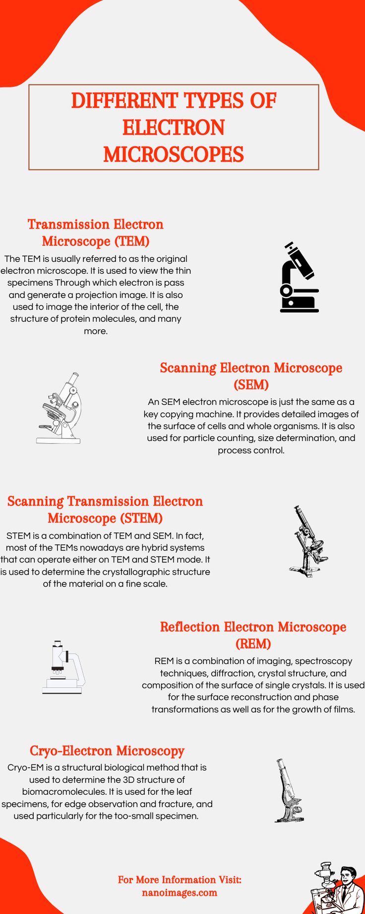
Different Type Of Electron Microscopes Electron Microscope Scanning Electron Microscope Electrons
0 Response to "If You Viewed One Single Protein Using a Microscope"
Post a Comment